All Cats Celebrates Twenty Years!

P1020958

Cherry Blossom says stray kitties need extra love. Please donate to our Stray Cat Fund.

On Saturday July 20th, All Cats HealthCare Clinic celebrated twenty years of taking care of the feline residents of Gainesville and the surrounding area. Some of our clients drive well out of their way, passing many other veterinary hospitals on the way, and for that, we are grateful.

The day was marked by an Open House, with each of the technicians presenting information about an aspect of the clinic in which they had a special interest or particular experience. Since I spent ten years Someplace Else doing veterinary radiography, I of course, made a modest Powerpoint for our clients, highlighting the radiologic capability we now have with Computed Radiography. So, may I present my humble creation.

RADIOLOGY open house presentation

Posted in Uncategorized | Leave a comment

Introduction to Veterinary Radiology

1. Introduction- Veterinary Radiology

The production of a radiograph involves the use of complicated apparatus and a sequence of complex physical processes. A basic understanding of radiological physics will allow the radiographer to make the best use of the equipment available.

RadioGraphy – the processes involved in getting a radiograph
RadioLogy – the procedure of looking at a radiograph

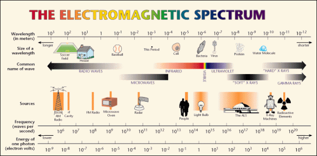
2. The Electromagnetic spectrum and ionising radiation

EMSpec

  1. X-Rays form only one part of the electromagnetic spectrum, which includes radio waves, microwaves (radar, heating etc.), infra-red, visible light, ultraviolet light, X-Rays and gamma rays.
  2. These radiations all have a few common properties:
    • They do not require a medium for propagation
    • They are transmitted by combined electric and magnetic fields
    • They travel with the same velocity in a vacuum
    • They travel in straight lines
    • They may interact with matter and be absorbed or scattered
    • They have the characteristics of both transverse waves and discrete particles photons)
  3. The energy of a photon is equal to the frequency multiplied by Planck’s constant
    • Thus photons of short wavelength have high frequency and high energy
    • These photons are useful in diagnostic radiology

 3. Properties of X-Rays

  1. Fluorescence
    • X-Rays cause certain substances to fluoresce, i.e. to emit light in the visible spectrum after absorbing X-rays
    • Such substances include calcium tungstate or rare-earth phosphors, both found in       intensifying screens (q.v.)
  2. Photographic effect
    • X-Rays produce a ‘latent image’ (invisible to the eye) on photographic film
    • This image is made visible by processing the film
  3. Penetration
    • X-Rays can penetrate substances or tissues that are opaque to visible light
    • They are gradually absorbed the further they pass through an object
    • The amount of absorption depends on the atomic number and density of the object and on the energy of the X-Rays
  4. Excitation and ionisation
    • X-Rays produce excitation and ionisation of the atoms and molecules of the substances through which they pass
    • Excitation is the process of raising an electron to a higher energy level
    • Ionisation is the process in which an electron is completely removed from an atom
  5. Biological effect
    • X-Rays interact with living tissue and can cause biological changes
    • These changes are mediated directly by excitation or ionisation of atoms or indirectly as a result of chemical changes occurring near the cells
    • Affected cells may be damaged or killed
    • Genetic effects involve chromosomal damage or mutation in the reproductive cells and will affect future generations
    • Somatic effects involve damage to the other tissues and result in changes within the       individual’s lifetime (e.g. radiation burns, leukemia)
    •  Radiation is a particular hazard because its effects are painless, latent and cumulative
    • SO QUIT COMPLAINING AND WEAR THE PROTECTIVE EQUIPMENT AND YOUR BADGE!!! 

4. The X-Ray tube

1. Since electrons are produced when fast-moving electrons are slowed down or stopped, there are three basic requirements for X-Ray production in an X-Ray tube:

  • A source of electrons (cathode)
  • A target to stop the electrons (anode)
  • A method of accelerating the electrons from the source to the target (high potential       difference between the two)
  1. The cathode:
    • Electrons are produced by heating a filament by thermionic emission
    • The number of electrons which pass from the cathode to the anode represents the tube current and is measured in milliamperes (mA)
    • The tube current can be controlled by the radiographer
  2. The anode
    • The anode is  maintained at a high positive potential with respect to the cathode       during the exposure
    • Electrons emitted by the filament are therefore accelerated to the anode and strike the target
    • The potential difference between the cathode and the anode is measured in kilovolts       (kV), and can be controlled by the radiographer
    • X-Rays are produced at the anode when it is struck by electrons; but only 1% of the incident energy is converted to X-Rays. The remainder is converted into heat, and       must be dissipated
    • A fixed anode relies on conduction of the heat away from the target by mounting it on a copper stem
    • A rotating anode allows X-Rays to be produced over a greater area of the target by       rotating it during the exposure

 5. Factors affecting the quality and intensity of beam

  1. Quality is a measurement of the penetrating power of the X-Ray photons. The quality of the beam increases as the proportion of high energy photons increases
  2. Intensity is a measure  of the amount of radiation produced
  3. Various factors affect the quality and intensity of the beam
    • kV – the greater the potential difference across the tube, the faster the electrons move and the higher the energy of the X-Ray photons. Thus the quality and       intensity are increased
    • mA – the higher the tube current, the greater the intensities of all the photon energies, and the intensity of the beam is increased
    • time – the longer the exposure, the greater the time during which X-Rays are produced, and the greater the beam intensity. The time (seconds) and mA are generally considered together as the composite factor mAs.  mA x time = mAs
    • distance – increasing distance from the source of radiation results in a decrease in the intensity of the beam, according to the inverse square law. Hence doubling the distance from the tube head will result in a beam of one quarter its original intensity

6. Interaction of X-Rays with matter

  1. When a beam of X-Rays passes through matter, its intensity is reduced as the energy is absorbed or scattered
  2.  The degree of attenuation depends on the atomic number and physical density of      the tissue, and the energy of the X-Rays
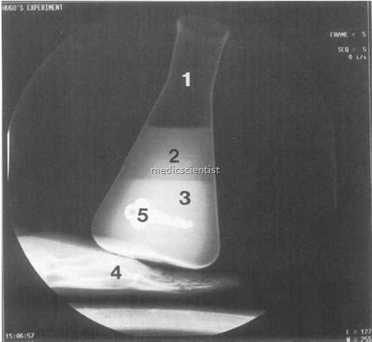
  3. The differential absorption of X-Rays by the various tissues of the patient allows a      radiographic image to be made

density


  1. 1. Air
  2. 2. Fat
  3. 3. SoftTissue
  4. 4. Bone
  5. 5. Metal


7. Properties of radiographic film and formation of the image

  1. X-Rays will produce a pattern of varying depths of blackness on the film
  2. The incident photons are attenuated to varying degrees by the patient’s tissues
  3. The emerging pattern of photons of differing energies affect the film emulsion to produce the latent image
  4. Developing the film allows this image to become visible
  5. The film emulsion consists of gelatin containing finely dispersed grains of silver bromide
  6. The absorption of X-Rays by the silver bromide grains renders them developable
  7. Silver bromide grains which have been exposed to X-Rays will be turned black by the developer
  8. Unexposed grains are dissolved and removed and leave a white area on the film

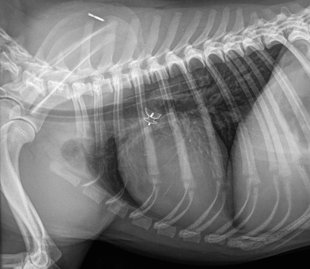
Consider the possibilities when making a radiograph of a patient’s thorax:

dog chest

  1. Some rays do not penetrate the chest at all, and are only slightly attenuated by air.. Therefore the maximum number of photons will pass through to the film. This causes maximum exposure of that part of the film, causing blackening of the film
  2. Some rays pass through the body wall and the lung. The body wall will absorb a number of photons, but few photons are absorbed by the aerated lung.
    Therefore the film will receive a medium exposure
  3. Some rays pass through the body wall, mediastinum and heart.
    Most of the photons will be absorbed, and only a few pass on to the film.
    This results in a low exposure of that part of the film, causing a relatively white area to be seen on the film

 8. Effect of the exposure factors on the film

  1. kV: governs the penetrating power of the photons
    • Films taken with a low kV have high contrast, i.e. tissues appear black or white with very little grey
    • Films taken with a high kV have low contrast, i.e. the film is more uniformly grey (a flat film)
  2. mAs: governs the amount of X-Rays reaching the film
    • mAs too low (underexposure) results in a pale film
    • mAs too high (overexposure) results in a dark film
  3. Distance: affects the amount of X-Rays reaching the film by the inverse square law
    • This will only affect  the film if the distance is changed without altering the mAs
  4. In essence, the kV  controls the number of shades of grey, and the mAs controls how dark they are

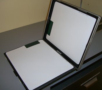
9. Cassettes and intensifying screens

xray_cassette_cervical_8x10_open

1. A cassette is a  light-tight, robust container which will:

  • Hold the film and intensifying screens in close contact
  • Protect the film from visible light, since this will blacken the film as well as X-Rays
  • Protect the film and screen from damage during use
  1. Two intensifying screens are present within the cassette, either side of the film
    • Screens contain materials which fluoresce when bombarded by X-Rays
    • The radiographic film is sensitive to this visible light emitted by the screens as well as the  X-Rays themselves
    • The use of both ]visible light and X-Ray photons allows the reduction of exposure times
    • However, film without screens or a cassette is used when very fine detail is required (Paper cassettes- require very long exposure times, usually a full second)

10. Qualities of a good diagnostic radiograph

  1. An accurate portrayal of the structures under examination, which requires good positioning with the minimum of geometric distortion (the region of interest should be  under the central ray, parallel to and close to the cassette)
  2. Easy perception of the relevant structures with good contrast and detail
  3. No misleading artifacts

11. Objective qualities of a good radiograph

  1. Contrast
    • This is the difference in blackness between two adjacent tones
    • Films with high contrast will show a white bone on a uniformly black background
    • Films with a low contrast will show no black or white, but only tones of grey

    Contrast is affected by

    • kV: low kV gives high contrast
    • mAs: low mAs gives low contrast
    • inherent properties of the radiographic film
    • processing: development affects contrast
    • scattered radiation produced within the patient reduces contrast
  2. fog:
    • inherent in the film
    • exposure to visible light or incorrect safe light in dark room
    • overdevelopment
  3. Detail and definition
    • The details of a radiograph depend for their perceptibility upon their contrast with the background and upon their definition or sharpness
    • Contrast has been dealt with above, however, it is important to note:
    • Very small details require high contrast if they are to be easily seen
    • If one opacity merges gradually into another, the eye will find it difficult to        perceive the difference
    • If two densities are  separated by a firm line of demarcation, the difference can easily be seen
    • Definition is poor if there is a blurred image, which may be caused by:
      • factors inherent in the tube head and film/screen combination
      • movement of the machine, animal or film during exposure

12. Identification of radiographs

  1. All films must be adequately labeled and show
    • The name of the hospital or practice
    • The date
    • The patient identity or case number
  2. Various methods are available
    • Lead letters
    • Lead-impregnated tape
    • Film camera: exposes the information into the emulsion
  3. Additional information may need to be incorporated
    • Left/right marker to identify limbs or radiographic projection
    • Indication of time elapsed in a sequential study
    • Pre- and  post-operative pictures

Radiographs are part of the medical record and MUST BE ADEQUATELY AND ACCURATELY LABELED.

 13. Radiation safety

ALARA is a philosophy of excellence used in one’s day-to-day work with radioactive materials and radiation sources. It is when one strives to keep one’s radiation exposure As Low As Reasonably Achievable.

Persons operating X-ray equipment should practice dose reduction methods whenever using the equipment.Basic safety measures include keeping exposure time short, keeping distance from the source large, and using appropriate shielding, such as lead aprons. All users of X-ray equipment must have specific training as provided by their supervisor.

Human fingers (or any other parts) should NEVER!!! be present in the radiograph.

Lead gloves do not provide protection from the primary X-ray beam. In other words, wearing a lead glove and putting your hand in the radiograph offers no protection at all. The lead only protects the hands from scatter radiation arising at the periphery of the radiographic image.

  1. Protection of the radiographer
    • Personnel should never be in the primary beam
    • Patients should not be held during an exposure unless it is clinically necessary, i.e. sedation or general anesthesia is required in many cases
    • All persons participating in the examination should wear appropriate protective       clothing (lead rubber aprons, gloves/sleeves)
    • Persons not involved in the radiographic examination should be excluded from the room
    • The room should be large and have protective screens for use during exposure
    • Appropriate dosimeters should be worn to identify if unacceptable exposure to X-Rays is occurring
  2. Protection of the patient
    • Use the fastest film/screen combination possible to obtain diagnostic pictures
    • Collimate the primary beam to include only the area of interest
    • Use a reasonable film-focal distance
    • Avoid repeat radiographs by ensuring proper exposure and development
  3. Protection of the public
    • Doors and walls  should be treated as required to prevent escape of radiation
    • Warning lights should be placed outside the radiography room to signal tube preparation and exposure
    • Radiation warning signs should be present on the doors
    • People under the age of 18 and pregnant women should not be allowed to assist in radiographic procedures

The majority of the text is from: http://www.priory.com/vet/physint.htm

Posted in Uncategorized | Leave a comment

Signs a cat may try to kill you.

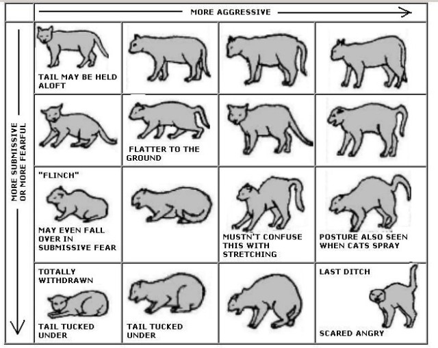
With our patients being exclusively feline, and new technicians not always having such intensive experience with cats, I thought I would post everything I could find that could help us read how a cat is feeling, so we can make the visit to the clinic as atraumatic as possible.

This is a general chart, the facial expressions going from benign in the upper left to full-on Screaming Mimi in the bottom left. I’m sure we’ve all seen examples of every one of these expressions, possibly on the same cat, sometimes within the space of just a few minutes.


But cats express themselves with more than just their face and eyes, right? So, the next chart shows the importance of tail position in helping us understand what a cat is feeling.

And this chart shows the whole package, including posture and body position.

For an in-depth discussion of cat body language, please follow this link to the website  from which I’ve stolen borrowed these graphics.

Posted in Uncategorized | Leave a comment

Commonly used medical terms

cardiomegaly– enlarged heart

caudal– toward the tail

dysuria– painful or difficult urination

ectropion– turning out of the eyelid (usually the lower eyelid) so that the inner surface is exposed, resulting in dry, painful eyes and excess tearing of the eye (epiphora);

edema– abnormal accumulation of fluid beneath the skin or in one or more cavities of the body that produces swelling.

entropion– turning in of the edges of the eyelid (usually the lower eyelid) so that the lashes rub against the eye surface.

epiphora– excess tearing of the eye

epistaxis– bleeding from the nose

erythema– redness of the skin

exopthalmia– protruding or bulging eyeballs

halitosis– foul-smelling breath

hematuria– blood in the urine

hyphema– Bleeding in the front portion of the eye

hypoalbuminemia– levels of albumin in blood serum are abnormally low.

hypocalcemia– low serum calcium levels in the blood

hypokalemia– lower-than-normal amount of potassium in the blood.

hyponatremia– a metabolic condition in which there is not enough sodium (salt) in the body fluids outside the cells.

iritis– a form of anterior uveitis and refers to the inflammation of the iris of the eye.

keratitis– a condition in which the eye’s cornea, the front part of the eye, becomes inflamed.

keratoconjunctivitis sicca (kcs)-(dry eye syndrome), is a relatively common inflammatory condition characterized by inadequate tear film protection of the cornea.

microcardia– abnormal smallness of the heart.

osteomyelitis– an infection of the bone or bone marrow

otitis– ear infection

panosteitis– a painful condition that affects the cat’s long leg bones and is characterized by limping and lameness.  It can occur with any breed, but it is more common in medium- to large-sized cat breeds and young cats around 5 to 18 months in age.

pododermatitis-inflammatory (red, hot, painful) condition of the feet.

pollakiuria– abnormally frequent urination

pruritis– itching

pthisis bulbi– is a shrunken, non-functional eye that results from severe ocular disease, inflammation, or injury.

pyelonephritis– an ascending urinary tract infection that has reached the pyelum or pelvis of the kidney.

pyorrhea– Purulent inflammation of the gums and tooth sockets, often leading to loosening of the teeth.

renomegaly– enlargement of the kidney(s).

rostral- toward the nose

splenomegaly– enlargement of the spleen

spondylosis– stiffening of the spine as a result of disease.

spondylitis– inflammation of the vertebra

stranguria– straining to urinate

trichobezoar– hairball

uveitis– swelling and irritation of the uvea, the middle layer of the eye.

Posted in Uncategorized | Leave a comment

Senior Cat Care Guidelines

The VetMed Team website is a great resource for continuing education for veterinary technicians. I am currently taking a free course there about senior cat care guidelines. Here is a link to a .pdf file that contains the Senior Cat Care Guidelines from 2008, from the American Association of Feline Practitioners.

Posted in Uncategorized | Leave a comment

Commonly used drugs and dosage calculations

Amlodipine– Antihypertensive- 2.5 mg tablets. Dosed at 0.625 mg

Ampicillin injectable– (125 mg/ml)- Antibiotic. Reconstitute with 7.5 ml of sterile water. Dosed at 10 mg/lb. Administered IV, TID. Expires in 24 hours. Given to dentals, 1 dose at induction, second dose two hours later.

Atropine

Baytril– (22.7 mg/ml) Antibiotic- Dosed at 5 mg/kg SID/injectable- give VERY slowly and dilute with saline- can cause blindness if given too quickly.

Buprenorphine– (o.3 mg/ml)- Analgesic. Dosed at 0.05-0.15 ml q 8-12 hours, depending on the patient’s size, for post-op pain. Administered sublingual (SL) or transmucosal (TM). Not effective if swallowed.

Cefazolin injectable – (100 mg/ml)- Antibiotic. Reconstitute with 10 ml of sterile water. Dosed at 10 mg/lb. Administered IV, first dose at induction, second dose two hours later. Given to spays/declaws/other surgeries.

Cerenia injectable- (maropitant citrate) – (10 mg/ml). Anti-nausea, antiemetic. Dosed at 1 mg/kg, administered SQ.

Cerenia tablets(maropitant citrate)- 16 mg tablets. Usually dosed at 1/4 tablet once daily for 3-4 days.

Clavomox liquid – (amoxicillin trihydrate/clavulanate potassium), 50 mg amoxicillin/12.5 mg clavulanate potassium. Antibiotic. Dosed orally BID. Can cause GI upset. Give on full stomach.

Clavomox tablets- (amoxicillin trihydrate/clavulanate potassium), 62.5 mg. Antibiotic. Dosed orally BID. Can cause GI upset. Give on full stomach.

Clindamycin– Antibiotic. Dosed BID, can be given SID depending on patient. Can cause GI upset. Give on full stomach. Can cause diarrhea.

Convenia injectable (Cefovecin sodium) (80 mg/ml)- Long-acting antibiotic. Dosed at 8 mg/kg. Administered SQ, left forelimb.

Diphenhydramine injectable– (trade name Benadryl, 50 mg.ml) Antihistamine. Used to premedicate against vaccine reactions, after insect stings, other allergic reactions. Give 15 minutes before administration of vaccine. Dosed at 0.5 mg/lb.

Emergency fluid rate= 10 ml/kg/hr. To calculate, take cat’s weight in pounds, divide by 2.2 to get kgs, multiply by 10, then divide result by 24 to get hourly rate.

Furosemide– (trade name Lasix, 50 mg/ml)- diuretic, used to treat hypertension. Dosed at 2-3 mg/kg

Mepron-Dosed at 15 mg/kg

Metronidazole injectable-(trade name Flagyl) (250 mg/ml) Dosed at 25 mg/kg.

Orbax (orbifloxacin) Antibiotic. Dosed at 3.4 mg/lb.

Panacur– (Fenbendazole)- (100 mg/ml): Broad spectrum anthelmintic (dewormer) Effective against roundworms, hookworms, whipworms, the taenia species of tapeworms, pinworms, aelurostrongylus, paragonimiasis, strongyles and strongyloides and can be administered to sheep, cattle, horses, fish, dogs, cats, rabbits and seals. Dosed at 50 mg/kg.

Prednisolone– Steriod- Dosage depends on patients and condition being treated. Can cause excessive thirst and/or hunger. Long term use can cause diabetes.

Ranitidine (trade name Zantac) -(25 mg/ml)- inhibits stomach acid production. Dosed at 2.5 mg/kg.

Posted in Uncategorized | 2 Comments

Labwork in Review (or: What do all these values mean?!)

We draw blood and collect urine so we can run these tests and determine the health status of our feline patients, but what do all those initials and values mean? Our lovely head technician Vanessa has compiled this overview to help us understand what all those letters and numbers mean.

Liver Values: (ALT, AST, ALKP, TBILI, GGT,CHOL, proteins)

  1. 1. ALT- Alanine aminotransferase is an enzyme found in liver cells. Damaged liver cells leak this enzyme which translates to increased quantities in the blood. This is a specific indicator of liver disease. It can be elevated without the cat being ill, if the thyroid level is high.
  2. AST- Aspirate aminotransferase is an enzyme found in many cells in the body, including the liver, heart and muscles.
  3. ALKP- Alkaline phosphatase is an enzyme present in the liver, bone, intestine or kidneys. Increases related to the liver can be caused by a blocked bile duct, actions of some drugs (such as steriods or phenobrabitol) or Cushing’s disease (hyperadrenocorticism). It can also be elevated in growing youngsters.
  4. TBILI (Total Bilirubin)- Bilirubin is a bile pigment that may be elevated in the blood if bile flow is obstructed or if the patient has Immune Mediated Hemolytic Anemia (IMHA) or any kind of anemia that involves red blood cell destruction (such as mycoplasma infection). It can be elevated without any other values being elevated if the cat has pancreatitis. It is often elevated in cats with liver disease and is the reason that icteric cats are yellow.
  5. ALBUMIN- Albumin is a plasma protein that is one half of the total protein level we check as part of the PCV/TP. A low albumin level can cause death if left untreated.
  6. GLOBULIN- is a plasma protein that is the other half of the TP reading. Elevations in globulins can be due to immune response, oral disease and illnesses like Feline Infectious Peritonitis (FIP).

Kidney Values: (BUN, CREA, PHOS, AMYL, ALB, URINARY SG, MICROALBUMIN, ELECTROLYTES)

  1. BUN (Blood Urea Nitrogen)- is made by the liver as a breakdown of dietary proteins related to ammonia. Increased BUN levels are indicative of kidney disease, decreased circulation to the kidneys and/or urinary  obstruction. If the BUN is elevated and the CREA is normal, it is possible that there is a GI bleed or, less likely, intestinal parasites.
  2. CREA (Creatinine)- is a by-product of phosphocreatine, a molecule involved in energy production in the skeletal muscles. It is basically a measure of permanent kidney damage. Keep in mind that elevation in the BUN/CREA levels isn’t seen until 75% or greater of the kidney function has been lost.
  3. Phosphorus- This is a by-product of food breakdown that is excreted by the kidneys. It is generally increased in cats with chronic renal dysfunction.
  4. Potassium- is a mineral that is essential for proper functioning of the heart, muscle, kidneys, nerves and digestive systems. Excessive elevations (hyperkalemia) in potassium can cause fatal cardiac arrythmias, as can hypokalemia. Cats with renal failure commonly present with hypokalemia due to urinary excretion of potassium. Additionally, cats with urinary blockages can have greatly increased levels of postassium.
  5. PCV (Packed red Cell Volume)- is a value that tells us whether or not the animal is anemic. Kitties that have chronic renal disease are always at risk of anemia because the kidneys excrete erythropoietin, the hormone that tells the bone marrow to produce red blood cells.
  6. Urinary Specific Gravity- This is a measure of how well the kidneys concentrate the urine. If the SG is below 1.035, it is an indicator that renal function is in some way compromised. HOWEVER, cats that eat a diet primarily of canned food or if they eat food with added water, the SG can be lower.
  7. Microalbumin- is a protein that is excreted in the urine. Experts are not sure if the protein causes worsening of the disease process or if the disease process causes the protein to be excreted. Whatever the reason, an elevated urine microalbumin means that the patient is far more likely to have a poor outcome to a medical challenge that an animal that doesn’t have it.

PANCREAS (SPEC FPL, GLUCOSE, AMYLASE, LIPASE)

  1. SPECIFIC FPL (feline pancreas-specific lipase)- This is a snap test that will  help diagnose or rule out pancreatitis in cats without a specific number attached. A positive specific fPL is indicative of pancreatitis.
  2. GLUCOSE- this checks the level of sugar in the blood. Glucose values can be elevated in cats due to diabetes or stress. Stress elevations in the BG should not cause the value to measure much over 250 mg/dL. If the blood levels exceed this, it is possibly due to diabetes mellitus.
  3. AMYLASE & LIPASE- both of these enzymes are excreted by the pancreas for food digestion. Elevations in these levels are clinically insignificant in cats. In dogs, elevations in amylase and lipase are indicative of pancreatitis.
  4. FRUCTOSAMINE– Checking this level can help differentiate between diabetes and stress elevations in the BG levels. If the value is high, it means that the BG has been elevated for a period of time and the cat is likely diabetic, rather than just stressed. When monitoring fructosamine in diabetic cats, if the levels are less than 500, it means that the diabetes is well-controlled.

THYROID

  1. THYROID- Thyroxin is a hormone produced by the thyroid gland, and overproduction of this thyroid hormone causes hyperthyroidism in cats. Hyperthyroidism is caused by a (usually) benign tumor of the thyroid gland. The total T4 test measures the amount of thyroxin being released in the body at the time of the blood draw. The free T4 test is a measure of the thyroxin present over a period of time, so it measures how much thyroxin is circulating in the body. Hyperthyroidism causes the body to run at a higher basal metabolic rate and can cause high blood pressure, detached retinas, gastrointestinal issues, hair loss, hyperactivity, congestive heart failure, kidney failure and eventually death.

URINALYSIS (STRIP, SG, SEDIMENTATION)

  1. STRIP- The urine strip measures the pH (normal is 6.3- 6.5), blood, bilirubin, protein (cats ALWAYS have protein on the strip), glucose, ketones, urobilinogen, and nitrites.
  2. SPECIFIC GRAVITY- Urinary Specific Gravity- This is a measure of how well the kidneys concentrate the urine. If the SG is below 1.035, it is an indicator that renal function is in some way compromised. HOWEVER, cats that eat a diet primarily of canned food or if they eat food with added water, the SG can be lower.
  3. SEDIMENTATION- This is performed to confirm the presence of blood, white blood cells (WBC’s), bacteria, cells, casts, crystals, and/or urinary parasites (such as capillaria).

CBC: Differential

LEUKOCYTES (NEUTROPHILS, LYMPHOCYTES, MONOCYTES, EOSINOPHILS, BASOPHILS)

  1. Neutrophils- These cells are sent out in response to infection or inflammation. They are about twice the size of red blood cells. The nucleus stains dark purple and the cytoplasm stains pale pink. In normal animals a healthy neutrophil nucleus will have two to four lobes. Neutrophils should comprise about 60-65% of the WBC’s seen on a differential.
    Immature neutrophils are also known as bands and are a direct sign of infection. The nucleus on bands is usually horseshoe shaped, the ends larger and rounded, with no ‘pinching’ or segmentation.
    Hypermature neutrophils are also known as toxic neutrophils.

    Neutrophil

    Band

Toxic Neutrophil

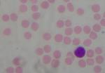
2.   Lymphocytes- These cells are usually seen as an acute response to a viral infection and are responsible for forming antibodies and antigenic response. The cell nucleus  stains purple with a thin band  of pale blue cytoplasm. In normal animals, the majority are mature, smaller cells that are 1.5 to 2 times the size of red blood cells. These should be approximately 30-40% of the cells seen.

Lymphocyte

3. Monocytes- these cells are normally a chronic response to infection, stress response, infectious processes or inflammation. On a CBC/Diff, about 8% monos may be seen on a normal animal.

Monocyte

4. EOSINOPHILS-  These cells are released in response to allergies, GI inflammation, or (usually) parasitism. Eosinophils are about the same size as neutrophils, or about twice the size of RBC’s.  The nucleus is lobulated or partially segmented and stains dark purple.  The cytoplasm stains a light blue color and contains red granules.  Feline eosinophils have small, rod-shaped granules. Maybe 2% of the leucocytes seen on a differential will be eosiniphils on a normal healthy cat.

Eosinophil

5. BASOPHILS- These cells are rarely seen in cats, but they are produced in response to severe allergies or severe parasitism.  Basophils are about the same size as neutrophils.  The nucleus is lobulated or partially segmented and often appears coiled.  The cytoplasm stains a blue to grey color and contains granules that vary in color from lavender to black.  The color and quantity of the granules vary with the species.   Feline eosinophils have numerous lavender granules.

basophil

An easy way to remember the correct proportions of the leucocytes in a differential (courtesy again of Vanessa) is to think of the phrase, “Never Let My Engine Blow, 60, 40, 8, 2, 0.” Yeah, I know, it doesn’t add up to 100%, but it gets the idea across.

THROMBOCYTES:

  1. PLATELETS- play a crucial role in the clotting of blood, along with all the clotting factors that are produced by the liver.

Platelets with a normal eosinophil
Courtesy of the Nottingham Vet School

ERYTHROCYTES

  1. RED BLOOD CELLS (RBC’s)- These are the cells that carry oxygen from the lungs to our tissues. Anemia (a deficiency in RBC’s) can be cause by blood loss, decreased production of new cells and/or RBC destruction. Anemia can be severe enough to result in death.

Top to bottom: 2 neutrophils with platelets below, lymphocyte, eosinophil, monocyte. All cells reported as unremarkable.

Posted in Uncategorized | Tagged , , , | 2 Comments

Greetings!

Welcome to the continuing story of an old dog (me) trying to learn new skills. My goal is to publish the educational materials provided to us at All Cats Healthcare Clinic for a couple reasons. Primarily, by passing this information through my brain by adding it here, I hope that more of it will stick. Also, by creating this blog, I hope to provide a useful online resource for the technicians at our clinic. I hope this will be useful.

Debi

Posted in Uncategorized | 2 Comments